
ProteinBlender: A New Tool for Molecular Animation
With funding from an NIGMS MIRA, the Iwasa lab is excited to embark on a new project to create new software tools for creating and sharing dynamic molecular visualizations!
Visit the project page to download ProteinBlender and learn how to use it!
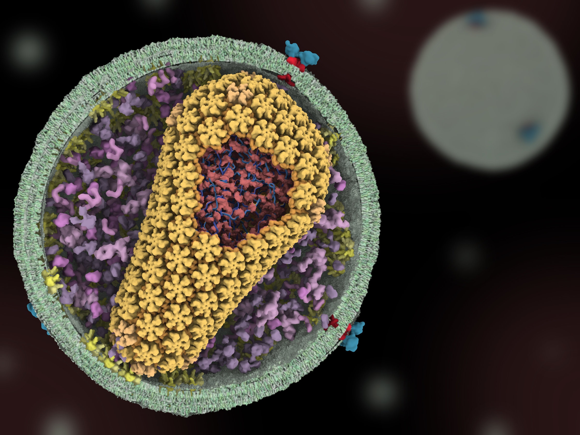
Science of HIV
Human immunodeficiency virus (HIV) is the causative agent of AIDS (Acquired Immunodeficiency Syndrome), a disease which was first described in the United States in the early 1980s. Since its initial discovery, HIV/AIDS has risen to become a global pandemic, with over 30 million infected individuals worldwide.
In collaboration with members of the CHEETAH consortium, this project seeks to describe current understanding of the molecular mechanisms by which HIV infects cells.
Please visit the Science of HIV website to view the animations and learn more about the project.

Water and Life Interface Institute (WALII)
The Animation Lab is happy to be a member of WALII! We are interested in visualizing the mechanisms by which some organisms, such as some plant seed, yeast, and tardigrades, are able to survive desiccation.

Cryo-EM 101
Cryo-EM has emerged as a powerful tool for high-resolution structure determination. To aid the training efforts of newcomers to the field, we are creating a media-rich curriculum to augment users’ own hands-on training. The training material will contain videos, animations, and interactive simulations that cover the major components of the cryo-EM workflow.
This project is a collaboration with Peter Shen.
PAST PROJECTS

Phase Separation 101
Recent research has shown that cells are organized not only through membrane-bound organelles, but also through liquid droplets assembled by Liquid–Liquid Phase Separation. This new paradigm, challenging the 20th century textbook view of cellular compartmentalization, is attracting growing attention as these biocondensates seem to play important roles in cells' physiopathology, and is now making its way into undergraduate classrooms.
This project aims at helping students understand the basics of this emerging, intricate and dynamic concept. To view the animations click here.

SARS-CoV-2 Life Cycle
In collaboration with the Visualization Design Lab at the University of Utah and support from the Coronavirus Structural Task Force and the National Science Foundation, we are creating an interactive and annotated visualization of the SARS-CoV-2 life cycle.
To learn more about the project and to see the animation click here.

Fat Metabolism
Completed by Shraddha Nayak with funding from the Utah Diabetes and Metabolism Research Center, this project describes how the body releases and stores fat, and how metabolic diseases like diabetes impact fat metabolism. Check out the lesson (and many accompanying animations): here. This project is funded by a grant from the Diabetes and Metabolism Research Center at the University of Utah.
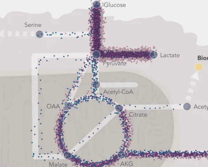
The Journey of a Metabolite
Completed by Shraddha Nayak with funding from the Huntsman Cancer Institute, this is a visual lesson on metabolic flux, describing glycolysis, the TCA cycle, and many other metabolic pathways. Check out the lesson (and many accompanying animations): here. This project is funded by a Pilot grant from the Huntsman Cancer Institute at the University of Utah.

Plant Cytokinesis
We are working with Georgia Drakakaki (UC Davis) to create an animation on plant cytokinesis. This project was funded by NSF and resulted in the following publication:
Plant cytokinesis and the construction of new cell wall.
Sinclair R, Hsu G, Davis D, Chang M, Rosquete M, Iwasa JH, Drakakaki G.
FEBS Lett. 2022 Sep;596(17):2243-2255. doi: 10.1002/1873-3468.14426. Epub 2022 Jul 15.

Transcriptional Regulation by Mediator
We are working together with the lab of Dylan Taatjes (UC Boulder) to create a series of animations that illustrate the role of Mediator in transcriptional regulation.
Check out some of the animations that Shraddha has completed with the Taatjes lab here!
The Mediator complex as a master regulator of transcription by RNA polymerase II.

Visualizing Tumor Metabolism
We are working together with the lab of Marcia Haigis (Harvard Medical School) to visualize different aspects of tumor metabolism and maintenance.

Modes of Vesicle Exocytosis
Flora Ye, a biology major from Fudan University, spent 6 months with the Animation Lab as an undergraduate intern. She researched and created an animation describing three different modes of vesicle exocytosis in neurons.
Read more about the project and see more of Flora’s work at her website here: https://www.flora-ye-visual.com. You can also view and download the animation here.

Visualizing Cdc48
In collaboration with the Shen lab at the University of Utah, we created an animation of the AAA+ ATPase Cdc48 in the process of translocating a substrate. Download the animation from Science here.
Structure of the Cdc48 segregase in the act of unfolding an authentic substrate.
Cooney I, Han H, Stewart MG, Carson RH, Hansen DT, Iwasa JH, Price JC, Hill CP, Shen PS.
Science. 2019 Aug 2;365(6452):502-505.

Molecular Flipbook
Molecular Flipbook was originally conceived as a prototype software that would allow biology researchers to readily create and share molecular animations. The project was funded by NSF and launched in 2014. You can watch Janet’s TED talk about it here, watch a tutorial video here, and read a short article about it here. The source code is available here.
Although this project is no longer active, we hope to integrate Flipbook-like functionality into other tools.

Centromeric Chromatin during Mitosis
In collaboration with Ben Black and postdoc Praveen Allu, we created an animation that depicts the structural transitions that centromeric nuclesome complexes undergo during mitosis.

Visualizing Vps4
In collaboration with the Hill lab at the University of Utah, we created an animation of the AAA+ ATPase Vps4 translocating a two-stranded substrate.
Structure of Vps4 with circular peptides and implications for translocation of two polypeptide chains by AAA+ ATPases.
Han H, Fulcher JM, Dandey VP, Iwasa JH, Sundquist WI, Kay MS, Shen PS, Hill CP.
Elife. 2019 Jun 11;8. pii: e44071. doi: 10.7554/eLife.44071.

Optic Cup Illustration
We created a 3D illustration of an optic cup, highlighting the release of RNA-filled exosomes, to describe recent results from the lab of Stephen Redenti.

Chromatin Remodeling
In collaboration with Brad Cairns and Cedric Clapier (Huntsman Cancer Institute, University of Utah and HHMI), we created a series of animations describing how chromatin is remodeled by different complexes, including ISWI and SWI/SNF.
These animations were published as supplemental figures in a review and can be viewed and downloaded here.

Architecture of the Type IV Pilus Machine
Using a grappling-hook like mechanism, some bacteria are able to attach to a substrate and pull themselves forward. The molecular machinery and the mechanism for this type of motility was animated for this project, which was a collaboration with Yi-wei Chang in Grant Jensen's group (Caltech).
Click here to view the abstract, and here to view the animated model on Youtube.
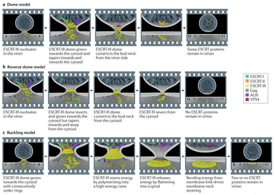
Membrane Scission by the ESCRT Proteins
In this collaboration with Jim Hurley's lab (UC Berkeley) and with support from CHEETAH, a series of three animations were constructed that depicted different models for how ESCRT III mediates membrane fission. Click on the links to view and play animations of the Dome model, Reverse Dome model, and the Buckling model or the publication abstract.

Cell Image Library
The Cell Image Library is a repository for images and movies of cells from a variety of organisms. It demonstrates cellular architecture and functions with high quality images, videos, and animations. This comprehensive and easily accessible Library is designed as a public resource for research and as a tool for education.
The Cell Image Library was initially funded and launched as an initiative by the American Society for Cell Biology.

Exploring Origins
The goal of the Exploring Origins project is to use molecular illustration and animation to help describe origins of life research and theories to broad audiences.
This website was part of a multimedia exhibit at the Museum of Science that included live presentations on the Current Science & Technology stage and a touch-screen kiosk.
Animations and illustrations were made during a NSF Discovery Corps Postdoctoral Fellowship, in collaboration with Jack Szostak and his laboratory at Massachusetts General Hospital, and the Current Science and Technology team at the Museum of Science.